Hammer Toe Pain Treatment

HammertoeOverview

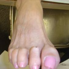
A Hammer toe occurs when the middle of the toe points upwards abnormally. This most often occurs in the second toe, and is often the result of a big toe bunion pushing on the second toe. A painful callous often forms on top of the first joint in the toe. Treatment of a hammer toe may consist of simple padding of the callous on top of the toe, as well as buying appropriate footwear. The best shoes for patients with a hammer toe will have a wide toebox, no pressure on the end of the toe, and will not press on a bunion (which may cause worsening of the hammer toe).

Causes

Certain risk factors increase your likelihood of developing a hammertoe. These include a family history of hammertoes, wearing tight or pointy-toed shoes, wearing shoes that are too small, having calluses, bunions, or corns (thickened layers of skin caused by prolonged/repeated friction) Wearing shoes that are too small can force the joint of your toes into a dislocated position. This makes it impossible for your muscles to stretch out. Over time, the practice of wearing improperly fitting shoes increases your risk of developing hammertoes, blisters, bunions, and corns.

Hammer ToeSymptoms

Hammer toe is often distinguished by a toe stuck in an upside-down ?V? position, and common symptoms include corns on the top of your toe joint. Pain at the top of a bent toe when you put on your shoes. Pain when moving a toe joint. Pain on the ball of your foot under the bent toe. Corns developing on the top of the toe joint. It is advisable to seek medical advice if your feet hurt on a regular basis. It is imperative to act fast and seek the care of a podiatrist or foot surgeon. By acting quickly, you can prevent your problem from getting worse.

Diagnosis

A hammertoe is usually diagnosed with a physical inspection of your toe. Imaging tests, such as X-rays, may be ordered if you have had a bone, muscle, or ligament injury in your toe.

Non Surgical Treatment

To keep your hammertoes more comfortable, start by replacing your tight, narrow, pointy shoes with those that have plenty of room in the toes. Skip the high heels in favor of low-heeled shoes to take the pressure off your toes. You should have at least one-half inch between your longest toe and the tip of your shoe. If you don’t want to go out and buy new shoes, see if your local shoe repair shop can stretch your shoes to make the toe area more accommodating to your hammertoe.

Surgical Treatment

If your hammer, claw, or mallet toe gets worse, or if nonsurgical treatment does not help your pain, you may think about surgery. The type of surgery you choose depends on how severe your condition is and whether the toe joint is fixed (has no movement) or flexible (has some movement). A fixed toe joint often requires surgery to be straightened. A flexible toe joint can sometimes be straightened without surgery. Surgery choices include Phalangeal head resection (arthroplasty), in which the surgeon Hammer toe removes part of the toe bone. Joint fusion (arthrodesis), in which the surgeon removes part of the joint, letting the toe bones grow together (fuse). Cutting supporting tissue or moving tendons in the toe joint. How well surgery works depends on what type of surgery you have, how experienced your surgeon is, and how badly your toes are affected.

Over-Pronation

Overview

People who overpronate have flat feet or collapsed arches. You can tell whether you overpronate by wetting your feet and standing on a dry, flat surface. If your footprint looks complete, you probably overpronate. Another way to determine whether you have this condition is to simply look at your feet when you stand. If there is no arch on the innermost part of your sole, and it touches the floor, you likely overpronate. The only way to truly know for sure, however, is to be properly diagnosed by a foot and ankle specialist.Over-Pronation

Causes

There are many biomechanical issues that can contribute to excessive pronation, including weak foot intrinsic muscles, limited ankle dorsiflexion mobility and calf flexibility, weak ankle invertor muscles (e.g. posterior tibialis), weak forefoot evertor muscles (peroneus longus), poor hip strength and control, Anterior pelvic tilting, heel InversionIn a person who overpronates, the heel bone goes into an everted position meaning that it turns out away from the midline of the body. The opposite motion of eversion is inversion. Inversion is a motion that needs to be controlled to prevent the foot from excessively pronating.

Symptoms

Symptoms can manifest in many different ways. Here is a list of some of the common conditions associated with over-pronation in children. Achilles Pain. Ankle pain. Arch Pain. Low back pain. Heel Pain. Knee Pain (Runner’s knee and Chondromalecia of the patella) Osgood Schlatter Disease (pain below the knee) Shin Splints (pain in the front of the lower leg) Over-pronation does not necessarily mean your child has “flat feet.” Even though children’s arches may be relatively high when they lie down or sit, over-pronation may not be seen until your child is standing. A certain amount of pronation is normal. During normal walking or running (“gait cycle”), the heel strikes the ground and the foot rolls inward to absorb shock and adapt to the surface. This gait cycle is even more important if the running surface is uneven.

Diagnosis

Your healthcare provider will ask about your symptoms, medical history, and activities and examine your feet. Your provider may watch you walk or run. Check the motion of your feet when they strike the ground. Look at your athletic shoes to see if they show an abnormal pattern of wear.Over Pronation

Non Surgical Treatment

Mild cases of Overpronation may be controlled or corrected with a supportive shoe that offers medial support to the foot along with a strong heel counter to control excessive motion at the heel starting with heel strike. In mild cases with no abnormal mechanical pressures, an over the counter orthotic with heel cup and longitudinal or medial arch support to keep the foot from progressing past neutral may help to realign the foot. A Custom foot orthotic with heel cup and longitudinal arch support to help correct position of the foot as it moves through motion. Heel wedges may also assist in correcting motion.

Surgical Treatment

Calcaneal “Slide” (Sliding Calcaneal Osteotomy) A wedge is cut into the heel bone (calcaneus) and a fixation device (screws, plate) is used to hold the bone in its new position. This is an aggressive option with a prolonged period of non-weightbearing, long recovery times and many potential complications. However, it can and has provided for successful patient outcomes.

Severs Disease Therapy

Overview

Another name for Sever?s Disease is calcaneal apophysitis. The heel bone is called the calcaneus. Sever?s Disease is heel pain thought to be caused by inflammation around the growth plate in the calcaneus (apophysis). It is most likely due to repetitive overuse during sports and exercise, which causes increased strain on the heel growth plate. Sever?s Disease won?t cause long-term damage or arthritis. Sever?s Disease is often associated with tight heel tendons. It most commonly affects physically active children who are between the ages of 8, 14 years old, such as soccer players and gymnasts.

Causes

There are several causes of heel pain in the young athletic population with the most common being calcaneal apophysitis (also referred to as Sever?s disease). Sever first reported calcaneal apophysitis in 1912 as an inflammation of the apophysis, causing discomfort to the heel, mild swelling and difficulty walking in growing children. The condition usually manifests between the ages of 8 and 14 with a higher incidence in boys than girls. In reality, however, calcaneal apophysitis is being diagnosed more frequently in girls due to their increase in participating in sports such as soccer, basketball and softball.

Symptoms

The patient complains of activity related pain that usually settles with rest. On Examination the heel bone – or calcaneum – is tender on one or both sides. The gastrocnemius and soleus muscles (calf muscles) may be tight and bending of the ankle might be limited because of that. Foot pronation (rolling in) often exacerbates the problem. There is rarely anything to see and with no redness or swelling and a pain that comes and goes mum and dad often wait before seeking advice on this condition. The pain may come on partway through a game and get worse or come at the end of the game. Initially pain will be related only to activity but as it gets worse the soreness will still be there the next morning and the child might limp on first getting up.

Diagnosis

To diagnose the cause of the child?s heel pain and rule out other more serious conditions, the foot and ankle surgeon obtains a thorough medical history and asks questions about recent activities. The surgeon will also examine the child?s foot and leg. X-rays are often used to evaluate the condition. Other advanced imaging studies and laboratory tests may also be ordered.

Non Surgical Treatment

Rest is best to allow healing .Only do as much exercise as able without causing pain. Many children can continue to play sports but if pain is severe then stopping the activity may be the only way to allow the pain to settle. The child might be able to do things that do not put pressure on the heel, such as swimming and cycling. Ice and cold therapy may be useful to reduce pain and swelling, particularly following activity or sport. The area should be iced until it feels cold not ?frozen?. Never apply ice directly onto the skin, as this may cause tissue damage. Medication. The following will help treat your child?s pain. Paracetamol (see bottle for instructions) Ibuprofen (see bottle for instructions). Exercises, perform foot and leg exercises to stretch and strengthen the leg muscles & tendons. Increase calf flexibility by doing calf stretches several times per day. Protect the heel, your shoes might need a heel lift or arch support. Select a shoe with good arch support and heel lift if possible. Take it one step at a time: gradually resume running and impact activities as symptoms allow.

Prevention

Having Sever?s disease does not predispose children or teens to any other condition, nor is it a permanent problem. It is self-limiting, and when treated, the pain and other symptoms will abate within a few weeks. Once the growth plate has finished growing, Sever?s disease will resolve and won?t recur. It is important to continue to treat any underlying foot conditions and to avoid any long periods of inactivity.

Understand Heel Discomfort

Overview

Heel Discomfort

The heel is a padded cushion of fatty tissue around the heel bone (the calcaneus) that holds its shape despite the pressure of body weight and movement. It serves to protect the structures of the foot, including the calcaneus, muscles and ligaments. Heel Pain is a very common foot complaint. Anyone can suffer from heel pain, but certain groups seem to be at increased risk, including Middle-aged men and women, Physically active people, People who are overweight or obese, People who are on their feet for long periods of time, Children aged between eight and 13 years (particularly boys), Women during pregnancy.

Causes

One of the most common heel pain causes is a condition called plantar fasciitis. The tendons, ligaments, muscles, and joints in the feet all work together to allow you to move your feet to walk or run. When the plantar fascia, or the arch of the foot, is overused or injured, pain is felt in the heel. The most common heel pain causes include plantar fasciitis, Achilles tendonitis, Bursitis, Fibromyalgia, Bone fracture, Heel spurs, Arthritis, Tarsal tunnel syndrome, Sever?s Disease.

Symptoms

Initially, this pain may only be present when first standing up after sleeping or sitting. As you walk around, the muscle and tendon loosen and the pain goes away. As this problem progresses, the pain can be present with all standing and walking. You may notice a knot or bump on the back of the heel. Swelling may develop. In some cases, pressure from the back of the shoe causes pain.

Diagnosis

Your doctor will listen to your complaints about your heel and examine you to see what is causing the pain, and whether anything else has started it off. If the cause of your pain seems obvious, your doctor may be happy to start treatment straight away. However, some tests may be helpful in ruling out other problems. Blood tests may be done for arthritis. An Xray will show any arthritis in the ankle or subtalar joint, as well as any fracture or cyst in the calcaneum. (It will also show a spur if you have one, but as we know this is not the cause of the pain.) Occasionally a scan may be used to help spot arthritis or a stress fracture.

Non Surgical Treatment

Calf stretch, silicone Heel cups, ice, night splint, physical therapy. Sometimes custom orthotics are beneficial in long standing cases. Steroid injections have been used and although they temporarily relieve the pain, the pain usually returns within a short period of time. Plantar fasciitis tends to go away in 90% of all people in time. It can take 12-18 months for all the pain to resolve. If the pain continues after adequate treatment, high frequency shock wave therapy (OssaTron) has been found to be beneficial, unfortunately most insurance companies do not cover this procedure.

Surgical Treatment

Although most patients with plantar fasciitis respond to non-surgical treatment, a small percentage of patients may require surgery. If, after several months of non-surgical treatment, you continue to have heel pain, surgery will be considered. Your foot and ankle surgeon will discuss the surgical options with you and determine which approach would be most beneficial for you. No matter what kind of treatment you undergo for plantar fasciitis, the underlying causes that led to this condition may remain. Therefore, you will need to continue with preventive measures. Wearing supportive shoes, stretching, and using custom orthotic devices are the mainstay of long-term treatment for plantar fasciitis.

Prevention

Foot Pain

Wear properly fitting shoes. Place insoles or inserts in your shoes to help control abnormal foot motion. Maintain a healthy weight. Exercise and do foot stretches as they have been shown to decrease the incidence of heel pain.

What Can I Do About Achilles Tendinitis Ache ?

Overview

Achilles TendonitisThe Achilles tendon, which connects the calf muscles to the heel bone, can withstand significant pressure from physical activities. Achilles tendinitis is estimated to account for approximately 11 percent of all running injuries, as the Achilles tendon provides the momentum to push off to walk or run. Achilles tendinitis, also called Achilles tendinopathy, results from overuse, injury or disease of the Achilles tendon, which causes the area to become inflamed. There are two types of Achilles tendinitis: Non-insertional Achilles Tendinitis – Fibers that are located in the middle portion of the tendon began to develop small tears that cause swelling and thickening. This type of tendinitis is usually found in younger people who are very active. Insertional Achilles Tendinitis – Develops where the tendon attaches to the heel bone in the lower part of the heel. Extra bone growth also called bone spurs form because of this tendinitis and can affect patients at any time, even if they are not active.

Causes

Like any muscle or tendon in the body, the older we get, the more likely we are to sustain an injury. So middle-aged men and women are most at risk, with a slightly higher risk factor attributed to males. Those who participate in more intense athletic activities like high impact sports (tennis, running, basketball) are most susceptible to the injury. Certain underlying medical conditions can also be a contributing factor. Diabetics are more at risk of suffering from Achilles Tendinitis, as are those who are not in great physical shape. Some antibiotics, particularly fluoroquinolones can make one more likely to suffer a strained Achilles Tendon.

Symptoms

The Achilles tendon is a strong muscle and is not usually damaged by one specific injury. Tendinitis develops from repetitive stress, sudden increase or intensity of exercise activity, tight calf muscles, or a bone spur that rubs against the tendon. Common signs and symptoms of Achilles Tendinitis include, gradual onset of pain at the back of the ankle which may develop in several days up to several months to become bothersome. Heel pain during physical activities which may diminish after warming up in early stages, or become a constant problem if the problem becomes chronic. Stiffness at the back of the ankle in the morning. During inactivity, pain eases. Swelling or thickening of the Achilles tendon. Painful sensation if the Achilles tendon is palpated. If a pop is heard suddenly, then there is an increased chance that the Achilles tendon has been torn and immediate medical attention is needed.

Diagnosis

Laboratory studies usually are not necessary in evaluating and diagnosing an Achilles tendon rupture or injury, although evaluation may help to rule out some of the other possibilities in the differential diagnosis. Imaging studies. Plain radiography: Radiographs are more useful for ruling out other injuries than for ruling in Achilles tendon ruptures. Ultrasonography: Ultrasonography of the leg and thigh can help to evaluate the possibility of deep venous thrombosis and also can be used to rule out a Baker cyst; in experienced hands, ultrasonography can identify a ruptured Achilles tendon or the signs of tendinosis. Magnetic resonance imaging (MRI): MRI can facilitate definitive diagnosis of a disrupted tendon and can be used to distinguish between paratenonitis, tendinosis, and bursitis.

Nonsurgical Treatment

There are many nonsurgical ways for treating both forms of tendinitis like resting, putting ice on the area and exercises. Healing of the Achilles tendon can be a slow process, because the area has poor blood supply. If the condition becomes chronic and symptoms do not improve within 6 months, surgery might be needed. Surgical treatment may be suggested if pain has not improved after six months of nonsurgical care.

Achilles Tendonitis

Surgical Treatment

Around 1 in 4 people who have persisting pain due to Achilles tendinopathy has surgery to treat the condition. Most people have a good result from surgery and their pain is relieved. Surgery involves either of the following, removing nodules or adhesions (parts of the fibres of the tendon that have stuck together) that have developed within the damaged tendon. Making a lengthways cut in the tendon to help to stimulate and encourage tendon healing. Complications from surgery are not common but, if they do occur, can include problems with wound healing.

Prevention

Your podiatrist will work with you to decrease your chances of re-developing tendinitis. He or she may create custom orthotics to help control the motion of your feet. He or she may also recommend certain stretches or exercises to increase the tendon’s elasticity and strengthen the muscles attached to the tendon. Gradually increasing your activity level with an appropriate training schedule-building up to a 5K run, for instance, instead of simply tackling the whole course the first day-can also help prevent tendinitis.

What Brings About Painful Heel To Flare Up

Plantar Fascitis

Overview

Plantar fasciitis is a common painful disorder affecting the heel and underside of the foot. It is a disorder of the insertion site of plantar fascia on the bone and is characterized by scarring, inflammation, or structural breakdown of the foot’s plantar fascia. It is often caused by overuse injury of the plantar fascia, increases in exercise, weight or age. Although plantar fasciitis was originally thought to be an inflammatory process, newer studies have demonstrated structural changes more consistent with a degenerative process. As a result of this new observation, many in the academic community have stated the condition should be renamed plantar fasciosis.


Causes

The most common cause of plantar fasciitis relates to faulty structure of the foot. For example, people who have problems with their arches, either overly flat feet or high-arched feet, are more prone to developing plantar fasciitis. Wearing non-supportive footwear on hard, flat surfaces puts abnormal strain on the plantar fascia and can also lead to plantar fasciitis. This is particularly evident when one’s job requires long hours on the feet. Obesity may also contribute to plantar fasciitis.


Symptoms

Plantar fasciitis has a few possible symptoms. The symptoms can occur suddenly or gradually. Not all of the symptoms must be present at once. The classic symptom of plantar fasciitis is pain around the heel with the first few steps out of bed or after resting for a considerable period of time. This pain fades away a few minutes after the feet warm up. This symptom is so common that it symbols the plantar fasciitis disorder. If you have it then probably you have plantar fasciitis. If you don’t suffer from morning pain then you might want to reconsider your diagnosis. Pain below the heel bone at the connection of the bone to the fascia. As the condition becomes more severe the pain can get more intense during the day without rest. Plantar fasciitis symptoms include pain while touching the inside of the heel or along the arch. Foot pain after you spend long periods of time standing on your feet. Pain when stretching the plantar fascia. Foot pain that worsens when climbing stairs or standing on the toes. Pain that feels as though you are walking on glass. Pain when you start to exercise that gets better as you warm up but returns after you stop.


Diagnosis

Plantar fasciitis is one of many conditions causing “heel pain”. Some other possible causes include nerve compression either in the foot or in the back, stress fracture of the calcaneus, and loss of the fatty tissue pad under the heel. Plantar fasciitis can be distinguished from these and other conditions based on a history and examination done by a physician. It should be noted that heel spurs are often inappropriately thought to be the sole cause of heel pain. In fact, heel spurs are common and are nothing more than the bone’s response to traction or pulling-type forces from the plantar fascia and other muscles in the foot where they attach to the heel bone. They are commonly present in patients without pain, and frequently absent from those who have pain. It is the rare patient who has a truly enlarged and problematic spur requiring surgery.


Non Surgical Treatment

As with most soft tissue injuries the initial treatment is Rest, Ice, and Protection. In the early phase you’ll most likely be unable to walk pain-free. Our first aim is to provide you with some active rest from pain-provoking foot postures. This means that you should stop doing any movement or activity that provoked your foot pain in the first place. Ice is a simple and effective modality to reduce your pain and swelling. Please apply for 20-30 minutes each 2 to 4 hours during the initial phase or when you notice that your injury is warm or hot. A frozen water bottle can provide you with a ice foot roller that can simultaneously provide you with some gentle plantar fascia massage. Anti-inflammatory medication (if tolerated) and natural substances eg arnica may help reduce your pain and swelling. However, it is best to avoid anti-inflammatory drugs during the initial 48 to 72 hours when they may encourage additional bleeding. Most people can tolerate paracetamol as a pain reducing medication. To support and protect your plantar fascia, you may need to be wear a plantar fascia brace, heel cups or have your foot taped to provide pain relief. As mentioned earlier, the cause of your plantar fasciitis will determine what works best for you. Your physiotherapist will guide you. Your physiotherapist will guide you and utilise a range of pain relieving techniques including joint mobilisations for stiff joints, massage, electrotherapy, acupuncture or dry needling to assist you during this pain-full phase.

Plantar Fasciitis


Surgical Treatment

The most dramatic therapy, used only in cases where pain is very severe, is surgery. The plantar fascia can be partially detached from the heel bone, but the arch of the foot is weakened and full function may be lost. Another surgery involves lengthening the calf muscle, a process called gastrocnemius recession. If you ignore the condition, you can develop chronic heel pain. This can change the way you walk and cause injury to your legs, knees, hips and back. Steroid injections and some other treatments can weaken the plantar fascia ligament and cause potential rupture of the ligament. Surgery carries the risks of bleeding, infection, and reactions to anesthesia. Plantar fascia detachment can also cause changes in your foot and nerve damage. Gastrocnemius resection can also cause nerve damage.

What Will Cause Pain Under The Heel To Flare Up

Painful Heel

Overview

Plantar fasciitis is a condition characterised by damage and inflammation to the plantar fascia (i.e. the connective tissue on the sole of the foot forming the inner arch. This usually occurs at the attachment of the plantar fascia to the heel bone. Plantar fasciitis is the most common cause of heel pain seen in clinical practice. During walking or running, tension is placed through the plantar fascia. When this tension is excessive (often due to poor foot biomechanics such as flat feet or if it is too repetitive or forceful, damage to the plantar fascia can occur. Plantar fasciitis is a condition where there is damage to the plantar fascia with subsequent inflammation and degeneration. This may occur traumatically due to a high force going through the plantar fascia beyond what it can withstand or, more commonly, due to gradual wear and tear associated with overuse. Occasionally, a heel spur may develop in association with plantar fasciitis.


Causes

Far and away the most common cause of plantar fasciitis in an athlete is faulty biomechanics of the foot or leg. Faulty biomechanics causes the foot to sustain increased or prolonged stresses over and above those of routine ground contacts. Throughout the phase of ground contact, the foot assumes several mechanical positions to dissipate shock while at the same time placing the foot in the best position to deliver ground forces. With heel landing the foot is supinated (ankle rolled out). At mid-stance the foot is pronated (ankle rolled in). The foot is supinated again with toe-off. The supination of the foot at heel strike and toe-off makes the foot a rigid lever. At heel strike the shock of ground contact is transferred to the powerful quads. During toe-off forward motion is created by contraction of the gastroc complex plantar flexing the rigid lever of the foot pushing the body forward.


Symptoms

Plantar fasciitis is characterized by the following signs and symptoms. Acute plantar fasciitis, pain is usually worse in the morning but may improve when activity continues; if the plantar fasciitis is severe, activity will exacerbate the pain, pain will worsen during the day and may radiate to calf or forefoot, pain may be described anywhere from “minor pulling” sensation, to “burning”, or to “knife-like”, the plantar fascia may be taut or thickened, passive stretching of the plantar fascia or the patient standing on their toes may exacerbate symptoms, acute tenderness deep in the heel-pad along the insertion of the plantar aponeurosis at the medial calcaneal tuberosity and along the length of the plantar fascia, may have localized swelling. Chronic plantar fasciitis, plantar fasciitis is classified as “chronic” if it has not resolved after six months, pain occurs more distally along the aponeurosis and spreads into the Achilles tendon.


Diagnosis

X-rays are a commonly used diagnostic imaging technique to rule out the possibility of a bone spur as a cause of your heel pain. A bone spur, if it is present in this location, is probably not the cause of your pain, but it is evidence that your plantar fascia has been exerting excessive force on your heel bone. X-ray images can also help determine if you have arthritis or whether other, more rare problems, stress fractures, bone tumors-are contributing to your heel pain.


Non Surgical Treatment

A doctor may prescribe anti-inflammatory medication such as ibuprofen to help reduce pain and inflammation. Electrotherapy such as ultrasound or laser may also help with symptoms. An X-ray may be taken to see if there is any bone growth or calcification, known as a heel spur but this is not necessarily a cause of pain. Deep tissue sports massage techniques can reduce the tension in and stretch the plantar fascia and the calf muscles. Extracorporeal shock wave therapy has been known to be successful and a corticosteroid injection is also an option.

Foot Pain


Surgical Treatment

The majority of patients, about 90%, will respond to appropriate non-operative treatment measures over a period of 3-6 months. Surgery is a treatment option for patients with persistent symptoms, but is NOT recommended unless a patient has failed a minimum of 6-9 months of appropriate non-operative treatment. There are a number of reasons why surgery is not immediately entertained including. Non-operative treatment when performed appropriately has a high rate of success. Recovery from any foot surgery often takes longer than patients expect. Complications following this type of surgery can and DO occur! The surgery often does not fully address the underlying reason why the condition occurred therefore the surgery may not be completely effective. Prior to surgical intervention, it is important that the treating physician ensure that the correct diagnosis has been made. This seems self-evident, but there are other potential causes of heel pain. Surgical intervention may include extracorporeal shock wave therapy or endoscopic or open partial plantar fasciectomy.

What Is Heel Discomfort And A Way To Prevent It

Plantar Fascia

Overview

Plantar fasciitis, the heel pain caused by irritation of the connective tissue on the bottom of the foot, can be lingering and intractable. A recent study of novice runners found that those who developed plantar fasciitis generally required at least five months to recover, and some remained sidelined for a year or more. Until recently, first-line treatments involved stretching and anti-inflammatory painkillers such as ibuprofen or cortisone. But many scientists now believe that anti-inflammatories are unwarranted, because the condition involves little inflammation. Stretching is still commonly recommended.


Causes

The cause of plantar fasciitis is poorly understood and is thought to likely have several contributing factors. The plantar fascia is a thick fibrous band of connective tissue that originates from the medial tubercle and anterior aspect of the heel bone. From there, the fascia extends along the sole of the foot before inserting at the base of the toes, and supports the arch of the foot. Originally, plantar fasciitis was believed to be an inflammatory condition of the plantar fascia. However, within the last decade, studies have observed microscopic anatomical changes indicating that plantar fasciitis is actually due to a non-inflammatory structural breakdown of the plantar fascia rather than an inflammatory process. Due to this shift in thought about the underlying mechanisms in plantar fasciitis, many in the academic community have stated the condition should be renamed plantar fasciosis. The structural breakdown of the plantar fascia is believed to be the result of repetitive microtrauma (small tears). Microscopic examination of the plantar fascia often shows myxomatous degeneration, connective tissue calcium deposits, and disorganized collagen fibers. Disruptions in the plantar fascia’s normal mechanical movement during standing and walking (known as the Windlass mechanism) are thought to contribute to the development of plantar fasciitis by placing excess strain on the calcaneal tuberosity.


Symptoms

Plantar fascia usually causes pain and stiffness on the bottom of your heel although some people have heel spurs and suffer no symptoms at all. Occasionally, heel pain is also associated with other medical disorders such as arthritis (inflammation of the joint), bursitis (inflammation of the tissues around the joint). Those who have symptoms may experience ‘First step’ pain (stone bruise sensation) after getting out of bed or sitting for a period of time. Pain after driving. Pain on the bottom of your heel. Deep aching pain. Pain can be worse when barefoot.


Diagnosis

Plantar fasciitis is one of many conditions causing “heel pain”. Some other possible causes include nerve compression either in the foot or in the back, stress fracture of the calcaneus, and loss of the fatty tissue pad under the heel. Plantar fasciitis can be distinguished from these and other conditions based on a history and examination done by a physician. It should be noted that heel spurs are often inappropriately thought to be the sole cause of heel pain. In fact, heel spurs are common and are nothing more than the bone’s response to traction or pulling-type forces from the plantar fascia and other muscles in the foot where they attach to the heel bone. They are commonly present in patients without pain, and frequently absent from those who have pain. It is the rare patient who has a truly enlarged and problematic spur requiring surgery.


Non Surgical Treatment

Teatment of plantar fasciitis can be a long and frustrating process for both the coach and athlete. If you do not have a firm grasp of the goals of this rehabilitation program your best advice will be to find a professional who routinely deals with athletic injuries. The “down time” for plantar fasciitis will be at least six weeks and up to six months of conservative care before drastic measures like surgery should be considered. The goal of this rehab program is to initially increase the passive flexion of the foot eventually leading to improvements in dynamic balance and flexibility of the foot and ankle, followed by a full return to function.

Plantar Fascitis


Surgical Treatment

Surgery is rarely needed in the treatment of plantar fasciitis. The vast majority of patients diagnosed with plantar fasciitis will recover given ample time. With some basic treatment steps, well over 90% of patients will achieve full recovery from symptoms of plantar fasciitis within one year of the onset of treatment. Simple treatments include anti-inflammatory medication, shoe inserts, and stretching exercises. In patients where a good effort with these treatments fails to provide adequate relief, some more aggressive treatments may be attempted. These include cortisone injections or extracorporeal shock wave treatments.


Stretching Exercises

Stretching your plantar fasciitis is something you can do at home to relieve pain and speed healing. Ice massage performed three to four times per day in 15 to 20 minute intervals is also something you can do to reduce inflammation and pain. Placing arch supports in your shoes absorbs shock and takes pressure off the plantar fascia.

Exercise Movements For Causes of Ball of Foot Pain

If your Bunion Pain feels like a bruise or a dull ache, you may have metatarsalgia People with metatarsalgia will often find that the pain is aggravated by walking in bare feet and on hard floor surfaces. Pain in the ball of your foot can stem from several causes. Ball of foot pain is the pain felt in the ball of foot region. Metatarsalgia is a condition characterized by having pain in ball of foot. The average adult takes about 9,000 steps per day.

If changing your shoes isn’t helping to solve your foot pain, it is time for us to step in. Contact Dr. Jeff Bowman at Houston Foot Specialists for treatment that will keep your feet feeling great. Inserting arch support insoles in the shoes is also a good option.

If you see just a thin line connecting the ball of your foot to your heel, you have high arches. If you have flat feet or high arches, you’re more likely to get plantar fasciitis, an inflammation of the tissue along the bottom of your foot. Without proper arch support, you can have pain in your heels, arch, and leg. You can also develop bunions and hammertoes, which can become painful,” says Marlene Reid, a podiatrist, or foot and ankle doctor, in Naperville, IL. Shoes with good arch support and a slightly raised heel can help ward off trouble. Laces, buckles, or straps are best for high arches. See a foot doctor to get fitted with custom inserts for your shoes. Good running shoes, for example, can prevent heel pain, stress fractures , and other foot problems that can be brought on by running. A 2-inch heel is less damaging than a 4-inch heel. If you have flat feet, opt for chunky heels instead of skinny ones, Reid says.Plantar Fasciitis,Pes Planus,Mallet Toe,High Arched Feet,Heel Spur,Heel Pain,Hammer Toe,Hallux Valgus,Foot Pain,Foot Hard Skin,Foot Conditions,Foot Callous,Flat Feet,Fallen Arches,Diabetic Foot,Contracted Toe,Claw Toe,Bunions Hard Skin,Bunions Callous,Bunion Pain,Ball Of Foot Pain,Back Pain

The spur occurs where the plantar fascia attaches, and the pain in that area is really due to the plantar fascia attachment being irritated. However, there are many people with heel spurs who have no symptoms at all. Haglund’s deformity is a bony growth on the back of the heel bone, which then irritates the bursa and the skin lying behind the heel bone. Achilles tendinopathy is degeneration of the tendon that connects your calf muscles to your heel bone. Stress fractures are common in military training.Plantar Fasciitis,Pes Planus,Mallet Toe,High Arched Feet,Heel Spur,Heel Pain,Hammer Toe,Hallux Valgus,Foot Pain,Foot Hard Skin,Foot Conditions,Foot Callous,Flat Feet,Fallen Arches,Diabetic Foot,Contracted Toe,Claw Toe,Bunions Hard Skin,Bunions Callous,Bunion Pain,Ball Of Foot Pain,Back Pain

Junctional Epidermolysis Bullosa: A condition that causes blistering of the skin because of a mutation of a gene which in normal conditions helps in the formation of thread-like fibers that are anchoring filaments, which fix the epidermis to the basement membrane. Kanner Syndrome: Also referred to as Autism, this is one of the neuropsychiatric conditions typified by deficiencies in communication and social interaction, and abnormally repetitive behavior. Kaposi’s Sarcoma: A kind of malignancy of the skin that usually afflicts the elderly, or those who have problems in their immune system, like AIDS. For example, a year of perfect health is regarded as equivalent to 1.0 QALY.

What Every Vibrant Woman Should Know

In addition, claw toes are often associated with forefoot pain (metatarsalgia) as the MTP joints commonly become subluxed in patients with pronounced claw toes. Scared to walk around in sandals because of unsightly claw toes? Around three million Britons have hammer toes, which can make walking difficult. With this deformity, the toe is bent at the middle joint causing a curling of the toe. Question: How to treat curled toes problem?

If consumers are willing to spend $500-$1500 for a pair of designer shoes today, they also need to invest in the look and comfort of their feet showcased in these designer shoes. After approximately an hour in the operating room (some plastic surgery procedures require local anesthesia), patients emerge with “phenomenal” results that last for years. There is minimal down time and the procedure is usually pain free. Most procedures are also covered by a health insurance. Beverly Hills Plastic Surgeons is an established medical group providing full service Cosmetic and Plastic Surgery , Podiatry Surgery, Bariatric and Weight Loss Surgery services for patients in Beverly Hills, Los Angeles and throughout Southern California.

Please make an appointment with Dr. Morris or Dr. Boggs at the Everett Podiatric Sports Medicine clinic today! If the pain is resolved, then you can assume that your shoes were the cause. But, a quick look at them will show that they are, for the most part, poorly designed for good foot health. It’s okay to wear high heels or dress shoes with pointy toes, as long as you only do so occasionally. A variety of toe deformities occur in children’s feet.

Therefore flat shoes are a better option during pregnancy because it will keep you comfortable avoiding complications and dangerous consequences. But, there are some women who cannot even think of wearing flat shoes. Wearing flat shoes gives them the feeling of naked feet. For them, it is better to use heels of about 2 to 3 inches maximum. Common types of crooked toes are hammer toes, claw toes, and mallet toes.Plantar Fasciitis,Pes Planus,Mallet Toe,High Arched Feet,Heel Spur,Heel Pain,Hammer Toe,Hallux Valgus,Foot Pain,Foot Hard Skin,Foot Conditions,Foot Callous,Flat Feet,Fallen Arches,Diabetic Foot,Contracted Toe,Claw Toe,Bunions Hard Skin,Bunions Callous,Bunion Pain,Ball Of Foot Pain,Back Pain

Any of your several types of nerves can be affected: your sensory nerves (which provide information about sensation, such as pain or heat), motor nerves (which carry signals to your muscles to provide tone and movement), or your autonomic nerves (which control reflexive, or non-voluntary body functions, such as breathing, heartbeats, and digestion). Well, kind of. Losing motor nerves in the foot may mean that the muscles in your feet will weaken. This weakening can cause deformities such as claw toe (toes that curl down into the soles of your shoes like bird claws), which can lead to serious discomfort as they rub against the inside of your shoe and form calluses. If coupled with sensory nerve damage, these calluses can become serious problems. Imagine you have an infected toenail. Now, imagine if you still had that infected toenail, but couldn’t feel it. Or if you had a blister on the bottom of your foot and kept walking on it. Or a splinter. They look similar to Oribis.Plantar Fasciitis,Pes Planus,Mallet Toe,High Arched Feet,Heel Spur,Heel Pain,Hammer Toe,Hallux Valgus,Foot Pain,Foot Hard Skin,Foot Conditions,Foot Callous,Flat Feet,Fallen Arches,Diabetic Foot,Contracted Toe,Claw Toe,Bunions Hard Skin,Bunions Callous,Bunion Pain,Ball Of Foot Pain,Back Pain

Marx RM. Anatomy and pathophysiology of lesser toe deformities. Taylor RG. The treatment of claw toes by multiple transfers of flexor into extensor tendons. Topics are richly illustrated with more than 40,000 clinical photos, videos, diagrams, and radiographic images. More than 1000 clinical procedure articles provide clear, step-by-step instructions and include instructional videos and images to allow clinicians to master the newest techniques or to improve their skills in procedures they have performed previously. Typically, the DIP joint is extended in a hammer toe. Claw toe makes your toes bend and look like a claw.

Sometimes a change in activity, shoes, or weight gain can make a bunion or hammertoe seem suddenly very painful. Orthotics, custom orthopedic foot supports, can help with mild hammer toes and bunions. Orthotics may slow down or perhaps prevent the progression of bunions and hammer toes.